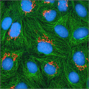
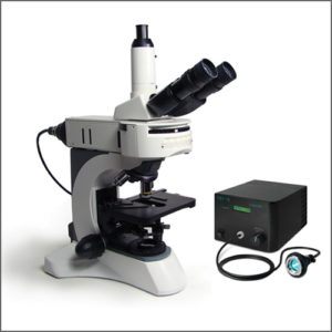


Fluorescence Microscope
Fluorescence microscope uses fluorescence and phosphorescence to study properties of organic or inorganic substances.
- Description
| Testing Method | Fluorescence Microscope |
| Description | Fluorescence microscope is an optical microscope that uses fluorescence and phosphorescence to study properties of organic or inorganic substances.
The specimen is illuminated with light of a specific wavelength (or wavelengths) which is absorbed by the fluorophores, causing them to emit light of longer wavelengths (i.e., of a different color than the absorbed light). The illumination light is separated from the much weaker emitted fluorescence through the use of a spectral emission filter. Typical components of a fluorescence microscope are a light source (xenon arc lamp or mercury-vapor lamp are common; more advanced forms are high-power LEDs and lasers), the excitation filter, the dichroic mirror (or dichroic beamsplitter), and the emission filter. The filters and the dichroic are chosen to match the spectral excitation and emission characteristics of the fluorophore used to label the specimen. In this manner, the distribution of a single fluorophore (color) is imaged at a time. Multi-color images of several types of fluorophores must be composed by combining several single-color images.
Most fluorescence microscopes in use are epifluorescence microscopes, where excitation of the fluorophore and detection of the fluorescence are done through the same light path (i.e. through the objective). These microscopes are widely used in biology and are the basis for more advanced microscope designs, such as the confocal microscope and the total internal reflection fluorescence microscope (TIRF).
In order for a sample to be suitable for fluorescence microscopy it must be fluorescent. There are several methods of creating a fluorescent sample; the main techniques are labelling with fluorescent stains or, in the case of biological samples, expression of a fluorescent protein. Alternatively the intrinsic fluorescence of a sample (i.e., autofluorescence) can be used. In the life sciences fluorescence microscopy is a powerful tool which allows the specific and sensitive staining of a specimen in order to detect the distribution of proteins or other molecules of interest. As a result, there is a diverse range of techniques for fluorescent staining of biological samples. |
| More Information | Wikipedia: Fluorescence Microscope |
Related Products
-
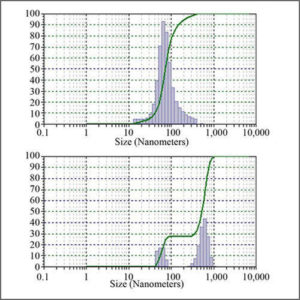
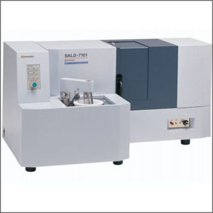
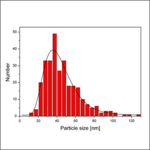
Particle Size Analyzer
Particle size analyzer provides accurate, reliable particle size distribution measurements from nanometers to millimetres.
-
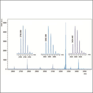
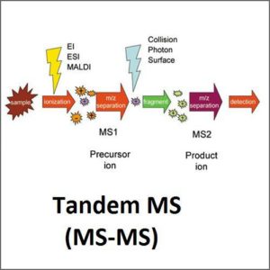
High Performance Liquid Chromatography – Mass Spectrometry (HPLC-MS)
High performance liquid chromatography – mass spectrometry (HPLC-MS) combines the physical separation capabilities of liquid chromatography with the mass analysis capabilities of mass spectrometry.
-
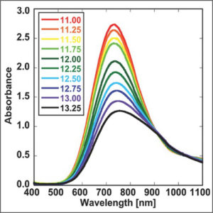
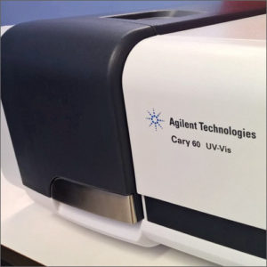
UV-Vis-NIR Spectroscopy
UV-Vis-NIR spectroscopy is routinely used in analytical chemistry for the quantitative determination of various analytes, such as transition metal ions, organic compounds, and biological macromolecules.
-
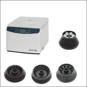
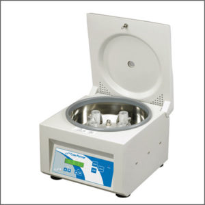
High-Speed Centrifuge
High-speed centrifuge is a method widely used to separate two immiscible substances involving the application of the centripetal force.
-
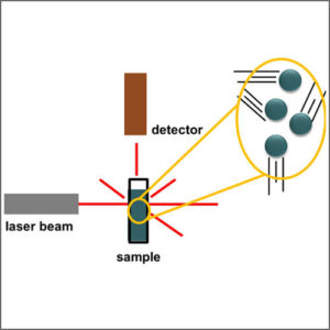
Laser Light Scattering (LLS)
Laser light scattering (LLS) is used to determine size of various particles including proteins, polymers, micelles and nanoparticles.