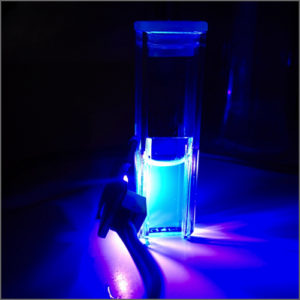


Fluorescence Spectroscopy
Fluorescence spectroscopy measures the fluorescent light emitted from a sample at different wavelengths, after illumination with a xenon flash lamp.
- Description
| Testing Method | Fluorescence Spectroscopy |
| Description |
Fluorescence spectroscopy is a type of electromagnetic spectroscopy that analyzes fluorescence from a sample. It involves using a beam of light, usually ultraviolet light, that excites the electrons in molecules of certain compounds and causes them to emit light; typically, but not necessarily, visible light. A complementary technique is absorption spectroscopy. In the special case of single molecule fluorescence spectroscopy, intensity fluctuations from the emitted light are measured from either single fluorophores, or pairs of fluorophores.
Molecules have various states referred to as energy levels. Fluorescence spectroscopy is primarily concerned with electronic and vibrational states. Generally, the species being examined has a ground electronic state (a low energy state) of interest, and an excited electronic state of higher energy. Within each of these electronic states are various vibrational states.
In fluorescence, the species is first excited, by absorbing a photon, from its ground electronic state to one of the various vibrational states in the excited electronic state. Collisions with other molecules cause the excited molecule to lose vibrational energy until it reaches the lowest vibrational state of the excited electronic state. This process is often visualized with a Jablonski diagram.
The molecule then drops down to one of the various vibrational levels of the ground electronic state again, emitting a photon in the process. As molecules may drop down into any of several vibrational levels in the ground state, the emitted photons will have different energies, and thus frequencies. Therefore, by analysing the different frequencies of light emitted in fluorescent spectroscopy, along with their relative intensities, the structure of the different vibrational levels can be determined.
For atomic species, the process is similar; however, since atomic species do not have vibrational energy levels, the emitted photons are often at the same wavelength as the incident radiation. This process of re-emitting the absorbed photon is “resonance fluorescence” and while it is characteristic of atomic fluorescence, is seen in molecular fluorescence as well.
In a typical fluorescence (emission) measurement, the excitation wavelength is fixed and the detection wavelength varies, while in fluorescence excitation measurement the detection wavelength is fixed and the excitation wavelength is varied across a region of interest. An emission map is measured by recording the emission spectra resulting from a range of excitation wavelengths and combining them all together. This is a three dimensional surface data set: emission intensity as a function of excitation and emission wavelengths, and is typically depicted as a contour map.
Fluorescence spectroscopy is used in, among others, biochemical, medical, and chemical research fields for analyzing organic compounds. There has also been a report of its use in differentiating malignant skin tumors from benign. |
| More Information | Wikipedia: Fluorescence spectroscopy |
Related Products
-
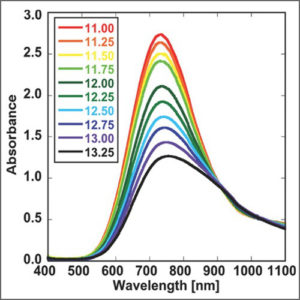
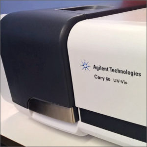
UV-Vis-NIR Spectroscopy
UV-Vis-NIR spectroscopy is routinely used in analytical chemistry for the quantitative determination of various analytes, such as transition metal ions, organic compounds, and biological macromolecules.
-
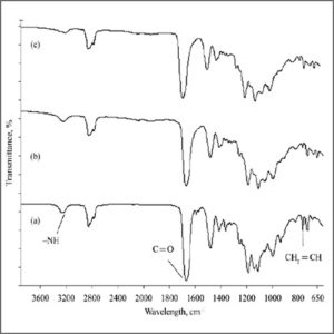
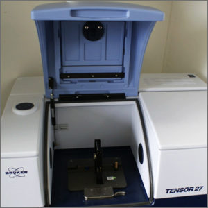
Fourier Transform Infrarred Spectroscopy (FTIR)
Fourier Transform-Infrared Spectroscopy (FTIR) is an analytical technique used to identify organic (and in some cases inorganic) materials.
-
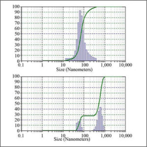
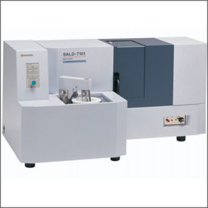
Particle Size Analyzer
Particle size analyzer provides accurate, reliable particle size distribution measurements from nanometers to millimetres.
-
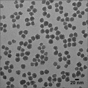
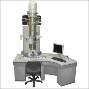
Transmission Electron Microscope (TEM)
Transmission electron microscope (TEM) utilizes energetic electrons to provide morphologic and size information on samples.
-
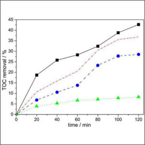
Total Organic Carbon Analyzer (TOC)
Total organic carbon analyzer (TOC) is used to determine the amount of carbon found in an organic compound.